 ANZBMS 2026
 Bilateral spontaneous atypical femoral fractures: a highly morbid presentation of adult hypophosphatasia after delayed diagnosis (#209)
Background Adult hypophosphatasia (HPP) is a rare genetic disorder caused by loss of function mutation in the ALPL gene on chromosome 1 encoding a tissue non-specific isoenzyme of alkaline phosphatase (TNSALP) (1). Accumulation of the enzyme’s natural substrate inorganic phosphate (PPi) leads to hypercalcaemia, hyperphosphataemia and hydroxyapatite growth inhibition.(1) The result is a low bone turnover state, intracellular B6 deficiency (elevated circulating B6), abnormal bone matrix (2) independent of bone mineral density and fractures. Inheritance can be autosomal dominant or recessive and diagnosis is often delayed but crucial to ensure appropriate management.
Case Presentation We present the case of a 74 year old retired non-smoker construction worker living independently home alone without significant supports. His comorbidities include gastric antral vascular ectasia, iron deficiency anaemia, hypertension and right knee replacement for osteoarthritis with no family history of skeletal dysplasia. His native dentition had been removed and replaced with dentures. He had unexplained low alkaline phosphatase (ALP) level in the fifteen years of available results prior to diagnosis and a humeral fracture post fall down two steps at age 67 years. There was no prior bisphosphonate or denosumab exposure.
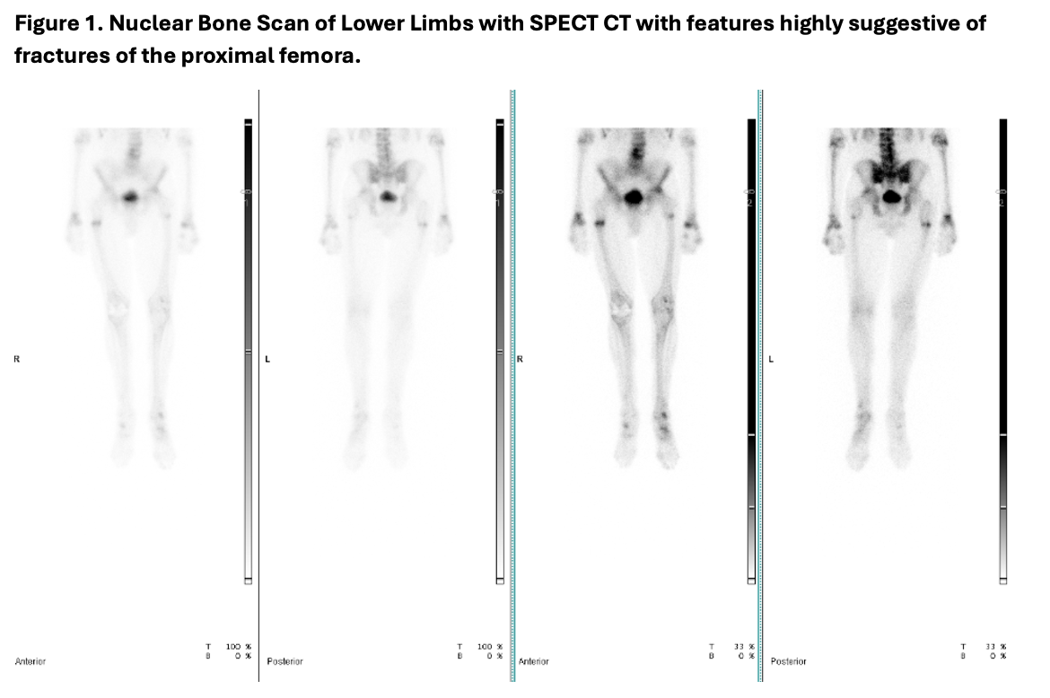
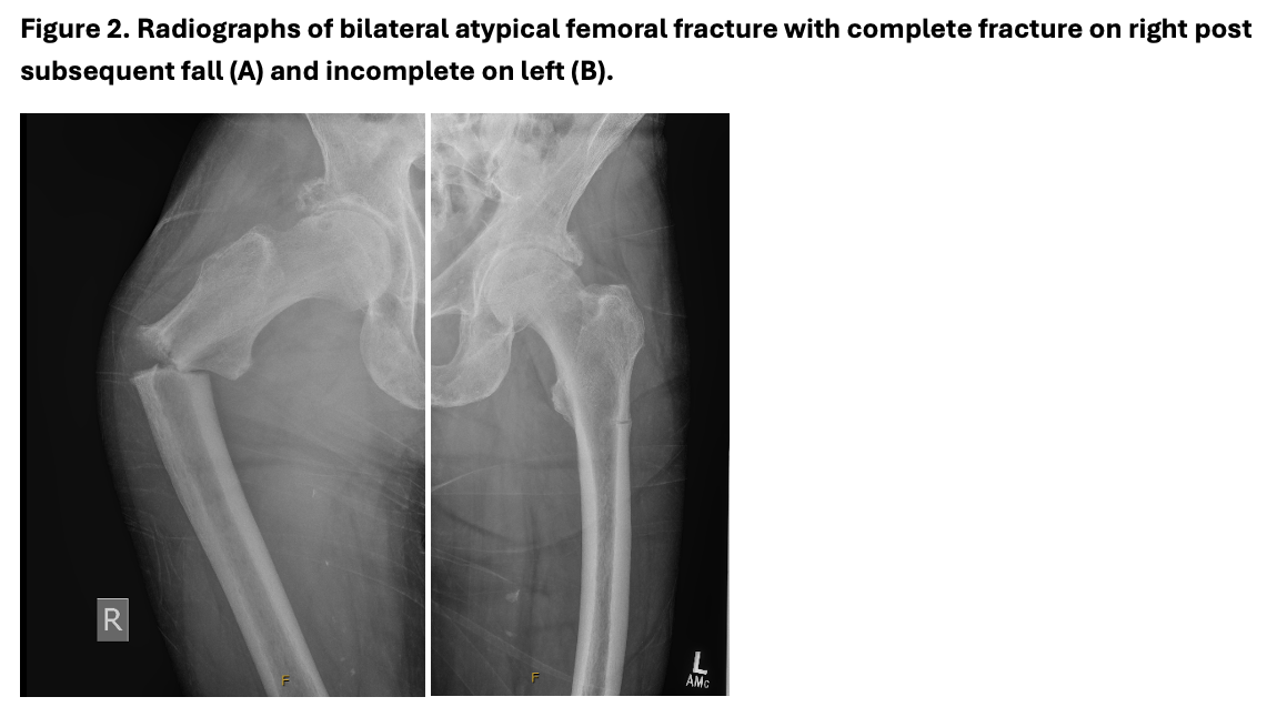
At aged 73 years, a bone scan (figure 1) conducted for right thigh and knee pain detected bilateral subtrochanteric incomplete femoral fractures (figure 2). After a minor fall, he sustained a complete right femoral fracture requiring surgical fixation and underwent subsequent prophylactic contralateral fixation.
Supportive of the underlying diagnosis was an elevated B6 840 nmol/L (reference 20-190). Investigations excluded thyroid disease, hyperparathyroidism, hypocalcaemia, zinc deficiency, hypomagnesaemia, vitamin D deficiency and coeliac disease. His phosphate was 1.60 mmol/L (reference 0.8-1.5) and baseline eGFR 67 ml/min/1.73m2. Bone turnover markers approximately one month after fracture were CTX 650 ng/L (reference 100-750) and P1NP 153 ug/L (reference 15-115). X-ray thoracolumbar spine revealed mild to moderate old compression fractures of multiple mid-dorsal vertebral bodies. Bone mineral density T-score at the lumbar spine (L1-L4) 2.8 likely reflected the compression fractures and osteophytes on x-ray while the left femoral neck t-score was 0.2 and total left femur t-score -0.1.
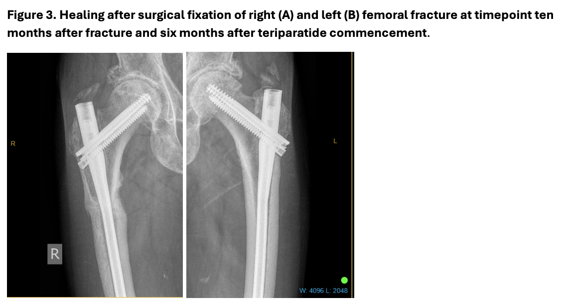
The post-operative course was complicated by debilitating pain, impaired mobility and significantly compromised quality of life and function. Hospital funded daily teriparatide 20mcg subcutaneously was commenced with a planned course of 12 months. Interval biochemical assessment after two months demonstrated CTX 1111 ng/L, P1NP 198 mcg/L, ALP 17 IU/L, eGFR 52 ml/min1.73m2, phosphate 1.64 mmol/L and corrected calcium 2.20 mmol/L (reference 2.1-2.6). Repeat pelvic X-ray confirmed bilateral satisfactory fracture union 6 months later. He had a full functional recovery without persistent pain and was able to continue living independently without support. Clinical genetics confirmation is awaited.
Case Discussion Timely diagnosis and investigation of low ALP levels is crucial in preventing the highly morbid and significant manifestations of adult hypophosphatasia. Even in patients with features before aged eighteen years, the mean diagnostic delay can be almost twenty-five years.(3) Atypical femoral fractures are a hallmark of this condition. They can be debilitating with associated pain, impaired mobility and protracted recovery. The typical presentation is of prodromal hip, groin or thigh pain and bilateral femoral pseudo-fractures in the lateral cortex of the subtrochanteric diaphysis.(1,4)
Pharmacological management of bone health in adult hypophosphatasia is challenging.(5) Unlike our case, adult HPP has been identified in those exposed to bisphosphonate therapy for suspected osteoporosis.(6) Bisphosphonates, analogues of inorganic pyrophosphate (PPi), can suppress bone turnover and deactivate TNSALP. Expert opinion is to avoid their use due to the risk of further impairing bone mineralisation, although the evidence is mixed.(7,8) Surgical management with load-sharing intramedullary fixation is the mainstay of management of atypical femoral fractures.
Case reports demonstrate teriparatide’s variable and often temporary improvement of ALP levels (9,10). Teriparatide may reduce fracture pain (11) and improve healing with an increase in the amount of osteoid and osteoblast numbers with enhanced bone formation(10,12). Our case exemplifies adequate fracture healing with teriparatide. The duration of therapy in the literature is typically 18 to 24 months, however alternate day administration and cyclical administration for longer periods may provide some benefit in bone mineral density and fracture prevention.(13)
Conclusions
- An unexplained low ALP and/or atypical femoral fracture without antiresorptive exposure requires timely investigation for a genetic cause such as adult hypophosphatasia to guide appropriate management.
- Atypical femoral fractures, particularly bilateral, are a hallmark of adult hypophosphatasia but can also been seen in other monogenic bone disorders.
- Unlike bisphosphonates, teriparatide may be useful to assist in fracture healing in patients with adult hypophosphatasia and prevent future fractures.
- 1. Whyte MP. Hypophosphatasia - aetiology, nosology, pathogenesis, diagnosis and treatment. Nat Rev Endocrinol. 2016 Apr;12(4):233–46. doi:10.1038/nrendo.2016.14 PubMed PMID: 26893260.
- 2. Zebaze R, Chiang CY, Shore-Lorenti C, Milat F, Ebeling PR. Disorganization of Bone Components in Metabolic and Genetic Bone Diseases like Hypophosphatasia: Novel Insights into the Pathogenesis and Patients Assessment. In. ASBMR; 2025 [cited 2026 Jan 15]. Available from: https://asbmr.confex.com/asbmr/2025/meetingapp.cgi/Paper/3916
- 3. Högler W, Langman C, Gomes da Silva H, Fang S, Linglart A, Ozono K, et al. Diagnostic delay is common among patients with hypophosphatasia: initial findings from a longitudinal, prospective, global registry. BMC Musculoskelet Disord. 2019 Feb 14;20:80. doi:10.1186/s12891-019-2420-8 PubMed PMID: 30764793; PubMed Central PMCID: PMC6376686.
- 4. Marini F, Brandi ML. Atypical femur fractures: a distinctive tract of adult hypophosphatasia. Clin Cases Miner Bone Metab. 2017;14(3):324–8. doi:10.11138/ccmbm/2017.14.3.324 PubMed PMID: 29354161; PubMed Central PMCID: PMC5762223.
- 5. Inoue D. Diagnosis and treatment of adult hypophosphatasia: Still a big challenge? Osteoporos Sarcopenia. 2024 Mar;10(1):1–2. doi:10.1016/j.afos.2024.03.002 PubMed PMID: 38690541; PubMed Central PMCID: PMC11056387.
- 6. Sutton RAL, Mumm S, Coburn SP, Ericson KL, Whyte MP. “Atypical femoral fractures” during bisphosphonate exposure in adult hypophosphatasia. J Bone Miner Res Off J Am Soc Bone Miner Res. 2012 May;27(5):987–94. doi:10.1002/jbmr.1565 PubMed PMID: 22322541.
- 7. Genest F, Seefried L. Subtrochanteric and diaphyseal femoral fractures in hypophosphatasia-not atypical at all. Osteoporos Int J Establ Result Coop Eur Found Osteoporos Natl Osteoporos Found USA. 2018 Aug;29(8):1815–25. doi:10.1007/s00198-018-4552-3 PubMed PMID: 29774402.
- 8. Rassie K, Dray M, Michigami T, Cundy T. Bisphosphonate Use and Fractures in Adults with Hypophosphatasia. JBMR Plus. 2019 Oct;3(10):e10223. doi:10.1002/jbm4.10223 PubMed PMID: 31687651; PubMed Central PMCID: PMC6820463.
- 9. Camacho PM, Mazhari AM, Wilczynski C, Kadanoff R, Mumm S, Whyte MP. Adult Hypophosphatasia Treated with Teriparatide: Report of 2 Patients and Review of the Literature. Endocr Pract. 2016 Aug 1;22(8):941–50. doi:10.4158/EP15890.OR
- 10. Gagnon C, Sims NA, Mumm S, McAuley SA, Jung C, Poulton IJ, et al. Lack of Sustained Response to Teriparatide in a Patient with Adult Hypophosphatasia. J Clin Endocrinol Metab. 2010 Mar 1;95(3):1007–12. doi:10.1210/jc.2009-1965
- 11. Whyte MP, Mumm S, Deal C. Adult Hypophosphatasia Treated with Teriparatide. J Clin Endocrinol Metab. 2007 Apr 1;92(4):1203–8. doi:10.1210/jc.2006-1902
- 12. Mizuno H, Sawa N, Sekine A, Inoue N, Oba Y, Ikuma D, et al. A Bone Histomorphometric Analysis of Hypophosphatasia-related Osteoporosis after Teriparatide Treatment. Intern Med. 2023;62(1):75–9. doi:10.2169/internalmedicine.9624-22
- 13. Polyzos SA, Tournis S, Goulas A, Kollia P, Whyte MP. Adult hypophosphatasia treated with reduced frequency of teriparatide dosing. J Musculoskelet Neuronal Interact. 2021;21(4):584–9. PubMed PMID: 34854399; PubMed Central PMCID: PMC8672398.