 ANZBMS 2026
 Breaking Convention: A Case Report of a Possible Atypical Ulna Fracture (#210)
Background
Prolonged antiresorptive therapy is associated with rare complications including atypical femoral fractures (AFF)1,2. A growing body of literature suggests the possibility of atypical fractures at non-femoral sites, such as the ulna3,4,5. We present the case of Mrs M, a 94-year-old woman who sustained bilateral AFFs, and a right proximal ulna fracture with possible atypical features, in the setting of prolonged antiresorptive treatment for osteoporosis.
Case Description
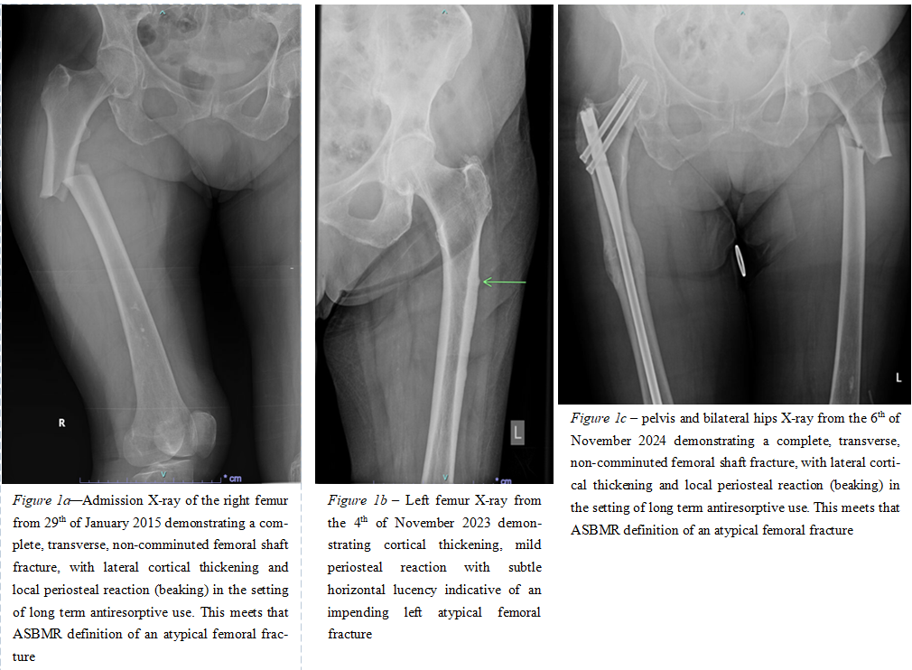
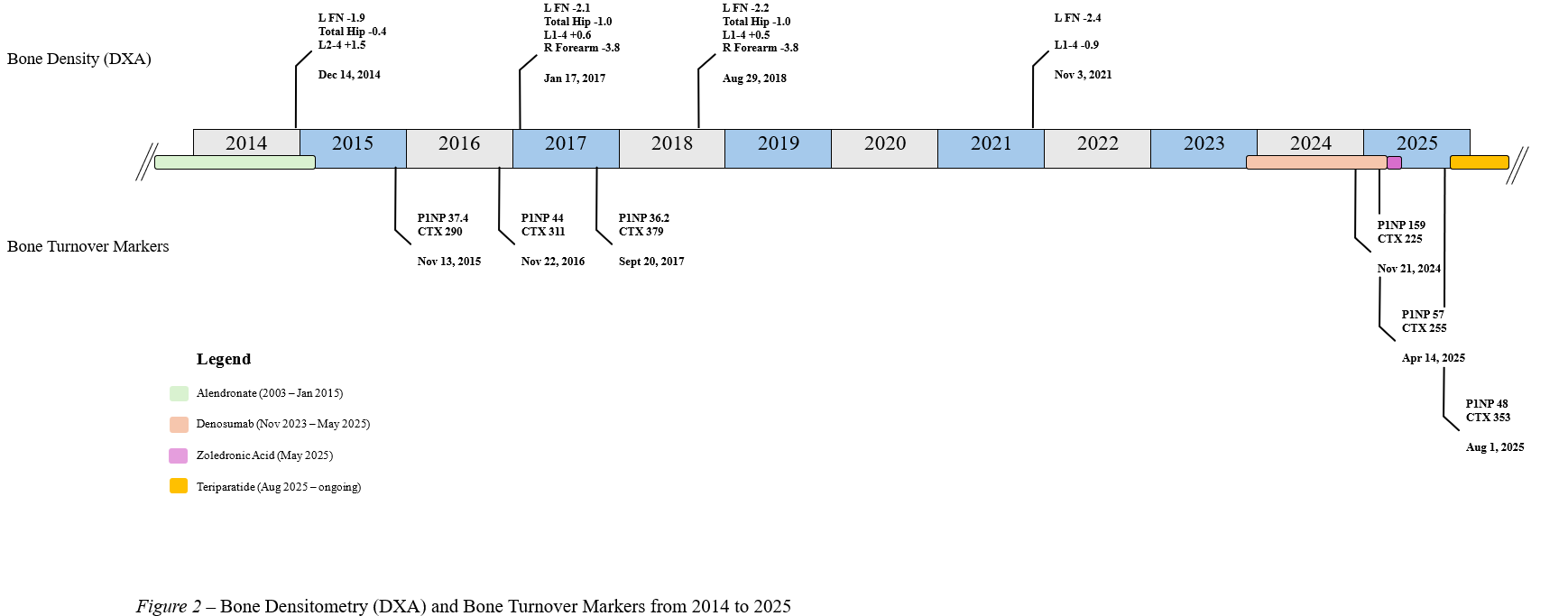
Mrs M was diagnosed with osteoporosis following a vertebral fracture in 2003 and commenced on alendronate 70mg weekly. This was continued uninterrupted until 2015 where, when rising from a chair, she sustained a right AFF (Figure 1a). This was managed with intramedullary (IM) femoral nail and alendronate ceased. At this time, Mrs M was living at home and mobilising with a 4-wheel walker. Her comorbidities included gastroesophageal reflux disease, diet-controlled type 2 diabetes mellitus, hypercholesterolaemia, hypertension, and breast cancer surgically cured 43 years prior. A screen for secondary causes of osteoporosis was unremarkable, and her bone density demonstrated osteopenia at the femoral neck (Figure 2).
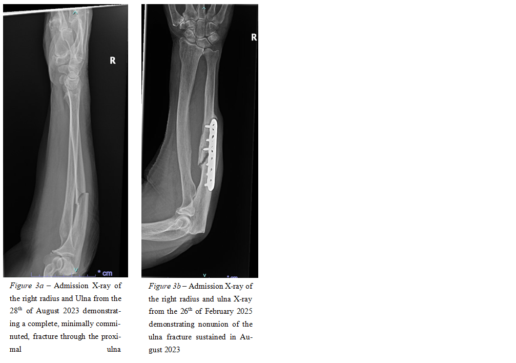
In November 2021, possibly due to persistent osteoporosis at the wrist (Figure 2), Mrs M’s GP commenced denosumab. In August 2023, following a fall from standing height, Mrs M sustained a right proximal ulna fracture (Figure 3a) without a history of prodromal pain. This was managed with ORIF using a locking plate. In November 2024, Mrs M fell whilst in the shower and sustained a left AFF (Figure 1c). Unfortunately, prodromal pain and lateral cortical beaking both present since August (Figure 1b) were not recognised as possible warnings of impending AFF. Her left AFF was managed with an IM femoral nail and denosumab was continued. She was readmitted in February 2025 with symptomatic malunion of her right ulna fracture (Figure 3b) requiring revision and bone graft application. In the context of prolonged antiresorptive exposure (cumulative 16 years), bilateral AFFs, poor healing and radiographic appearance of her ulna fracture - the diagnosis of atypical ulna fracture (AUF) was considered. To limit the exposure to antiresorptive therapy whilst balancing the risk of denosumab associated rebound bone loss, a decision was made to administer a single dose of zoledronic acid followed by a course of teriparatide. The first dose of zoledronic acid was administered in May 2025, and teriparatide was commenced in August 2025. A contralateral left forearm X-ray did not demonstrate radiological features concerning for impending AUF.
Case discussion
A recent systematic review by our group summarised the published literature describing antiresorptive-related atypical fractures at non-classical sites3. The most common site identified was the ulna. AUFs were found to share radiological features with AFF’s including transverse pattern, absence of comminution, and the presence of cortical beaking, best observed on lateral view of forearm radiographs3. Aside from the radiological appearance, the location of AUFs was consistently reported to occur at the posterior aspect of the proximal third of the shaft6. This is in contrast to fragility fractures of the forearm, which usually occurs at the distal forearm. Mrs M’s proximal ulna fracture (Figure 2a) meets four of the ASBMR’s five major criteria (minimal trauma; complete fracture; minimally comminuted; transverse pattern) and two minor criteria (delayed healing; associated bilateral AFF) thereby raising suspicion for a possible AUF.
Suppression of bone remodelling from prolonged bisphosphonate therapy results in altered intrinsic material properties of bone and impaired microdamage repair7,8. It is proposed that accumulation of microfractures may occur with prolonged antiresorptive therapy, and in response to repetitive forces, leads to an atypical fracture7,9,10,11. Atypical fractures are usually seen in the lateral aspect of the weightbearing femur, due to the greater biomechanical forces at this site, with femoral bowing being an additional risk factor. However, it is possible that antiresorptive therapy related atypical fractures can occur at non-femoral sites if the right local mechanical environment exists, such as using a walking aid which introduces repetitive bending motion on the forearm. Like the bending movement experienced by the femoral diaphysis due to the varus femorotibial angle12,13, we hypothesise that this may serve to concentrate tensile forces in the posterior cortex of the ulna thereby predisposing to AUFs.
Conclusion
There remains debate as to whether atypical fractures at non-femoral sites can occur. Case reports and series of AUFs have been reported in the literature, with the use of a walking aid identified as a risk factor in those with a predisposition to atypical fractures. Our case supports the hypothesis that AUFs may occur given the right intrinsic (bone) and extrinsic (mechanical) conditions.
Figures
- 1. Shane E, Burr D, Ebeling PR, et al. Atypical subtrochanteric and diaphyseal femoral fractures: report of a task force of the American Society for Bone and Mineral Research. J Bone Miner Res. 2010;25(11):2267–2294. https://doi.org/10.1002/jbmr.253
- 2. Shane E, Burr D, Abrahamsen B, et al. Atypical subtrochanteric and diaphyseal femoral fractures: second report of a task force of the American Society for Bone and Mineral Research. J Bone Miner Res. 2014;29(1):1–23. https://doi.org/10.1002/jbmr.1998
- 3. Collins L, Ronan A, Hutcheon E, Ebeling PR, Grill V, Nguyen HH. Atypical fractures at non-classical sites associated with anti-resorptive therapy: a systematic review. J Bone Miner Res. 2024;39(12):1722–1734. https://doi.org/10.1093/jbmr/zjae159
- 4. De Cicco A, Toro G, Oliva F, Tarantino U, Schiavone Panni A, Maffulli N. Atypical periprosthetic femoral fractures of the hip: a PRISMA compliant systematic review. Injury. 2021;52(8): 2407–2414. https://doi.org/10.1016/j.injury.2021.03.042
- 5. Tan SHS, Saseendar S, Tan BHM, Pawaskar A, Kumar VP. Ulnar fractures with bisphosphonate therapy: a systematic review of published case reports. Osteoporos Int. 2015;26(2):421–429. https://doi.org/10.1007/s00198-014-2885-0
- 6. Heo YM, Park SE, Cha SM, Shin HD, Choi JK. Diagnostic Criteria and Treatment of Atypical Ulnar Fractures Associated With Prolonged Bisphosphonate Therapy: Multicenter Case Analysis. J Hand Surg Am. 2022 Sep;47(9):901.e1-901.e12. doi: 10.1016/j.jhsa.2021.08.001. Epub 2021 Sep 24. PMID: 34565638
- 7. Ettinger B, Burr DB, Ritchie RO. Proposed pathogenesis for atypical femoral fractures: Lessons from materials research. Bone. 2013;55(2):495-500. https://doi.org/10.1016/j.bone.2013.02.004
- 8. Brock GR, Chen JT, Ingraffea AR, et al. The effect of osteoporosis treatments on fatigue properties of cortical bone tissue. Bone Reports. 2015;2(NA):8-13. https://doi.org/10.1016/j.bonr.2014.10.004
- 9. Alwahhabi BK, Alsuwaine BA. Long-term use of bisphosphonates in osteoporosis. Saudi Med J. 2017;38(6):604–608. https://doi.org/10.15537/smj.2017.6.19793
- 10. Mashiba T, Hirano T, Turner CH, et al. Suppressed Bone Turnover by Bisphosphonates Increases Microdamage Accumulation and reduces some biochemical properties in Dog Rib. J Bone Miner Res. 2000;15(4):613-620. https://doi.org/10.1359/jbmr.2000.15.4.613
- 11. Mashiba T, Turner CH, Hirano T, et al. Effects of high-dose etidronate treatment on microdamage accumulation and biomechanical properties in beagle bone before occurrence of spontaneous fractures. Bone. 2001;29(3):271-278. https://doi.org/10.1016/s8756-3282(01)00575-0
- 12. Martelli S, Pivonka P, Ebeling PR. Femoral shaft strains during daily activities: Implications for atypical femoral fractures. Clinical Biomechanics. 2014;29(8):869-876. https://doi.org/10.1016/j.clinbiomech.2014.08.001
- 13. Lo JC, Hui RL, Grimsrud CD, et al. The Association of Race/Ethnicity and Risk of Atypical Femur Fracture among Older Women Receiving Oral Bisphosphonate Therapy. Bone. 2016;85(NA):142-147. https://doi.org/10.1016/j.bone.2016.01.002