 ANZBMS 2026
 Automated Quantification of Osteoclast Fission Reveals Increased Osteoclast Fission Following Anti-RANKL Therapy (#229)
 Discontinuation of denosumab (Dmab), a monoclonal antibody targeting RANKL, causes a rebound increase in osteoclast formation and bone resorption that elevates fracture risk. Using an OPG:Fc withdrawal model, we previously identified an early rise in RANKL followed by a surge in osteoclast activity. Although accumulation of osteoclast precursors contributes to this phenomenon, the contribution of osteoclast fission and osteomorph formation remains unclear.
Our group recently identified osteomorphs, osteoclast-derived cells generated by fission that can rapidly re-fuse into functional osteoclasts. We hypothesised that anti-RANKL therapy promotes osteoclast fission, resulting in the accumulation of fusion-competent cells that contribute to rebound osteoclastogenesis following treatment cessation.
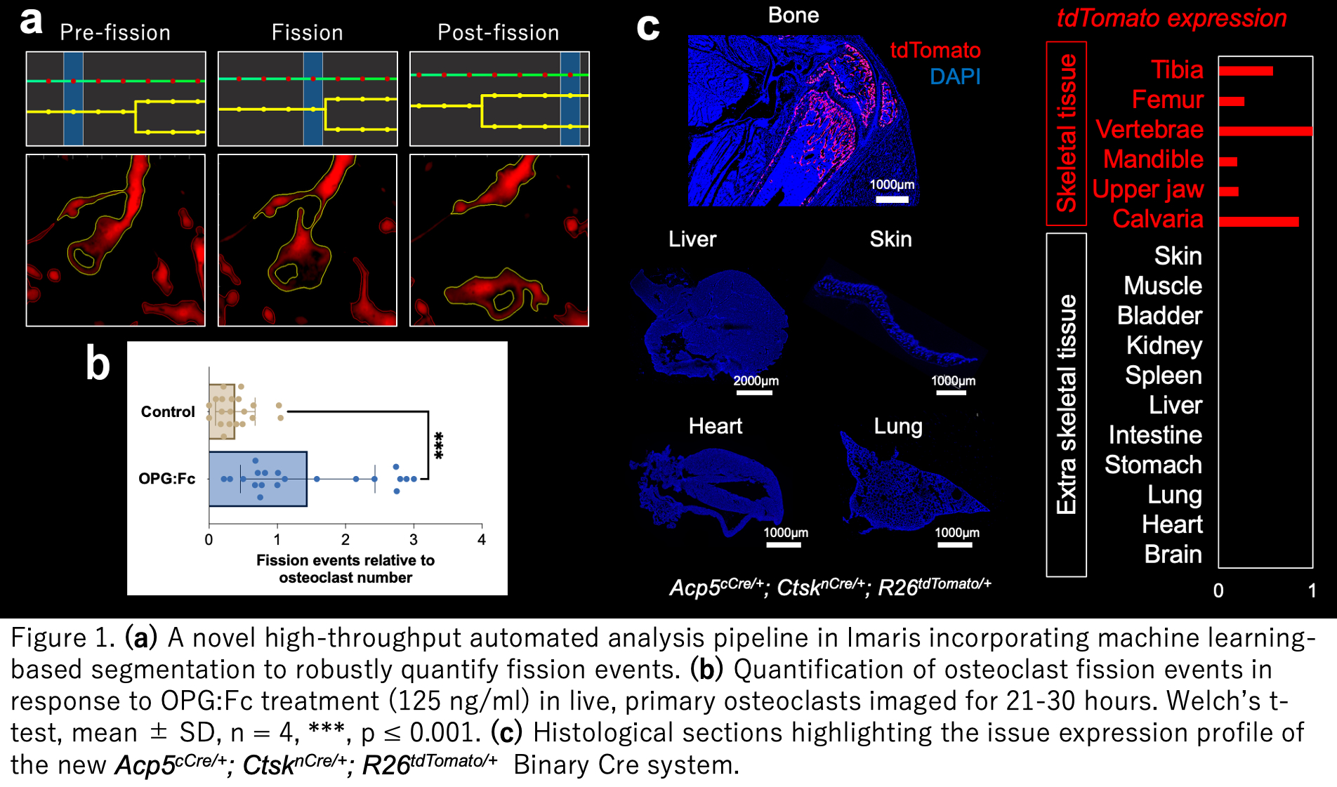
Using primary murine osteoclasts from LysM-Tdtomato mice and live-cell imaging, we previously demonstrated by manual analysis that low-dose OPG:Fc (31.25 ng/ml) significantly increased osteoclast fission. To evaluate the effects of RANKL inhibition on osteoclast dynamics in an unbiased manner, we developed a high-throughput automated analysis pipeline in Imaris incorporating machine learning-based segmentation to robustly quantify fission events (Fig. 1a). Using this approach, we identified a significant increase in osteoclast fission following treatment with high-dose OPG:Fc (125 ng/ml) compared with controls (Fig. 1b).
Ongoing studies will utilise a novel osteoclast-specific split-Cre reporter mouse driven by Acp5 and Ctsk promoters (developed by A/Prof Yahara’s laboratory) to enable precise tracking of osteomorphs and define their role in osteoclast reformation and Dmab rebound (Fig. 1c). Together, these findings implicate osteoclast plasticity as a driver of rebound bone loss and a potential therapeutic target in osteoporosis.