 ANZBMS 2026
 Atypical parathyroid adenoma presenting with severe PTH-dependent hypercalcaemia (#234)
Background: Primary hyperparathyroidism (PHPT) associated with severe hypercalcaemia, where serum calcium levels exceed 3.5 mmol/L, can be a potentially fatal condition without prompt management (1). Atypical parathyroid tumours (APTs) are responsible for approximately 1.3% of PHPT cases and are often difficult to distinguish pre-operatively from even rarer parathyroid carcinomas, presenting a diagnostic challenge (2). Diagnosis is based on histopathological assessment. We present a rare case of APT associated with profound PTH-dependent hypercalcaemia and review relevant diagnostic and follow-up considerations.
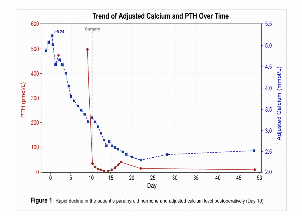
Case Presentation: A 56-year-old female presented with a four-day history of lethargy, anorexia and vomiting. Her medical comorbidities included type 2 diabetes mellitus and dyslipidaemia. Physical examination was notable for dry oral mucosa, with no confusion, bony tenderness or lymphadenopathy. Initial biochemistry revealed severe hypercalcaemia (corrected calcium of 4.89 mmol/L with a subsequent peak of >5.25 mmol/L) with a concurrent markedly elevated parathyroid hormone (PTH) of 454.3 pmol/L. Additional investigations included hypoalbuminaemia (28 g/L), hyperphosphataemia (1.85 mmol/L), elevated creatinine at 201 umol/L and low 25-hydroxyvitamin D at 16 nmol/L. Her troponin was elevated at 663 ng/L and electrocardiography revealed a shortened QT interval with upsloping lateral and inferior ST depression, reflective of demand ischaemia. Renal tract computed tomography showed no calculi.
Parathyroid ultrasonography identified a well-defined hypoechoic nodule posterior to the inferior pole of the right thyroid lobe measuring 10x6x10mm, consistent with parathyroid adenoma, and a 36mm TIRADS-3 right thyroid nodule. Computed tomography of the neck confirmed these findings. Treatment included aggressive intravenous hydration, two 30 mg doses of pamidronate, and salmon calcitonin 8 units/kg every eight hours for one week. Management was complicated by pulmonary oedema requiring loop diuretics and troponin leak; however, transthoracic echocardiography demonstrated preserved left ventricular function and no regional wall motion abnormalities. Following biochemical stabilisation to corrected calcium 3.01 mmol/L, she underwent right parathyroidectomy and hemithyroidectomy. Intraoperatively, a large right superior parathyroid adenoma was identified, while the right inferior gland appeared normal but was also removed due to clinical suspicion of carcinoma. The resected lesion measured 35×30×10mm and weighed 13g including thyroid tissue.
Histopathology demonstrated parathyroid tissue divided by thin fibrous bands, without marked pleomorphism or tumour necrosis. Immunohistochemistry showed retained parafibromin expression and a Ki-67 proliferation index <3%. Focal capsular invasion was present, with up to five mitoses/mm² and one atypical mitosis. Following specialist review, the lesion was classified as an APT rather than parathyroid carcinoma. Postoperatively, PTH fell promptly to 24.6 pmol/L. Recovery was uncomplicated, without hungry bone syndrome. Calcium supplementation was commenced, and outpatient review showed normalisation of calcium to 2.49 mmol/L with corresponding PTH 7.3 pmol/L at six weeks. The case was discussed at an endocrine multidisciplinary meeting, with consensus for six-month review using biochemical monitoring and neck ultrasonography. Genetic testing for CDC73 mutations is being considered. Her family history is unremarkable for related tumours.
Case Discussion: APTs are defined by atypical histological characteristics that raise concern for malignant potential but do not meet diagnostic criteria for carcinoma with local invasion or metastases (3). Reported features include fibrous bands, trabecular growth, pseudocapsular invasion, nuclear atypia, necrosis, intraoperative adherence and increased mitotic activity (4). However, no universally accepted threshold exists for the number or combination of features required for diagnosis.
Clinical and biochemical differentiation from benign adenoma or carcinoma also remains unreliable. Existing series suggest APTs may occur more commonly in women and at a younger median age of 44 years than typical benign disease, although presentations are heterogeneous (5). A low Ki-67 index and retained parafibromin may favour APT over carcinoma, but biomarker interpretation is limited by inconsistent reporting and small case numbers (5).
Longitudinal follow-up is important because recurrence, although uncommon (estimated up to 3%), has been reported many years after surgery (up to 17 years) and may be more frequent in hereditary disease, including germline CDC73-associated hyperparathyroidism-jaw tumour syndrome (5). No specific surveillance guidelines exist following parathyroidectomy. Practice varies by clinician experience, with some advocating for monitoring only for large-size tumours while others advise annual biochemical profile and neck ultrasound for 5 or 10 years to monitor for recurrence or development of carcinoma (6). Some authors suggest reserving genetic screening for patients with parafibromin loss or concerning family history (7).
Conclusions: This case highlights the diagnostic complexity and role of multidisciplinary management for APT presenting with profound hypercalcaemia. Histopathology remains the cornerstone of diagnosis whilst structured long-term follow-up is warranted, given rare recurrence and uncertain malignant potential. Further research is required to clarify recurrence risk, the role of genetic testing and establish evidence-based post-operative surveillance protocols.
- 1. Cetani F, Pardi E, Marcocci C. Update on parathyroid carcinoma. Journal of endocrinological investigation. 2016 Jun;39(6):595-606.
- 2. Galani A, Morandi R, Dimko M, Molfino S, Baronchelli C, Lai S, Gheza F, Cappelli C, Casella C. Atypical parathyroid adenoma: clinical and anatomical pathologic features. World Journal of Surgical Oncology. 2021 Jan 20;19(1):19.
- 3. DeLellis R, Larsson C, Arnold A, Lloyd R, Bilezikian J, Mete O, et al. Tumors of the parathyroid glands. In: Lloyd RV, Osamura RY, Klöppel G, Rosai J, editors. WHO Classification of Tumours of Endocrine Organs. 4th ed. Lyon: IARC Press; 2017. p. 157-70.
- 4. Rosai J, DeLellis R, Carcangiu M, Frable W, Tallin G. Parathyroid adenoma and variants. Tumors of the thyroid and parathyroid glands. Armed Forces Institute of Pathology; Atlas of tumor Patology 4th series. 2014:513-42.
- 5. Cetani F, Marcocci C, Torregrossa L, Pardi E. Atypical parathyroid adenomas: challenging lesions in the differential diagnosis of endocrine tumors. Endocrine-related cancer. 2019 Jul 1;26(7):R441-64.
- 6. Kumari N, Chaudhary N, Pradhan R, Agarwal A, Krishnani N. Role of histological criteria and immunohistochemical markers in predicting risk of malignancy in parathyroid neoplasms. Endocrine pathology. 2016 Jun;27(2):87-96.
- 7. Gill AJ, Lim G, Cheung VK, Andrici J, Perry-Keene JL, Paik J, Sioson L, Clarkson A, Sheen A, Luxford C, Elston MS. Parafibromin-deficient (HPT-JT type, CDC73 mutated) parathyroid tumors demonstrate distinctive morphologic features. The American journal of surgical pathology. 2019 Jan 1;43(1):35-46.