 ANZBMS 2026
 A diagnostic dilemma: a case report of PTH-independent hypercalcaemia in pregnancy (#32)
Background
Hypercalcaemia in pregnancy is an uncommon but clinically significant disorder, with potential maternal and foetal complications. Most cases are mediated by parathyroid hormone (PTH) with primary hyperparathyroidism being the most common aetiology. PTH-independent causes, are both rare and diagnostically challenging due to overlapping physiological changes in calcium and vitamin D metabolism during pregnancy. We describe a case of self-limited, PTH-independent hypercalcaemia of pregnancy, possibly due to an underlying impairment of vitamin D metabolism.
Case presentation
We report the case of AR, a 34-year-old primigravid woman evaluated for hypercalcaemia detected in late pregnancy, which was further complicated by diagnoses of hypertension in pregnancy and gestational diabetes mellitus. The patient’s past medical history included post-traumatic stress disorder, depression and a previous episode of urosepsis with subsequent acute kidney injury; she had no known history of hypercalcaemia or symptoms of hypercalcaemia. Her family history was significant for two biological siblings reportedly having recurrent episodes of nephrolithiasis in her birth country of Nepal. There was no known personal or family history of neuroendocrine tumours or other malignancy.
AR developed progressive hypercalcaemia [peak corrected calcium 3.02 mmol/L (2.15-2.65)) with a suppressed PTH [<0.3 pmol/L (2.0-8.5)], an undetectable PTH-related protein (PTHrP) [<0.1 pmol/L (<1.40)], normal 25-hydroxyvitamin D [79 pmol/L (>50)] and an inappropriately high-normal 1,25-dihydroxyvitamin D [1,25(OH)2D3] [183 pmol/L (50-190)]. Supplements during pregnancy included calcium carbonate 600 mg daily and vitamin D 7000 IU daily, commenced at 18 weeks’ gestation for pre-eclampsia prophylaxis. These were promptly ceased upon recognition of the patient’s hypercalcaemia, to little effect. Further investigations pointed away from multiple myeloma or granulomatous disease. Urinary calcium excretion was 5.7 mmol/24 hours (2.0-7.5), and urinary calcium clearance to creatinine clearance ratio was 0.7 (>0.01), arguing against familial hypocalciuric hypercalcaemia.
Despite intravenous hydration, hypercalcaemia persisted antenatally. Definitive management was attempted with delivery at 38+5 weeks, after which serum calcium rapidly normalised, with sustained normocalcaemia post-partum (Figure 1).
Repeat investigations six-months post-partum revealed low 25-hydroxyvitamin D (38 nmol/L), normal 1,25(OH)2D3 (91 pmol/L), and normal PTH (5.7 pmol/L). Urinary calcium excretion post-birth was 3.1 mmol/24 hours. A renal tract ultrasound demonstrated no renal calculi or nephrocalcinosis. The patient’s renal function remained robust with an eGFR of >90 mL/min/1.73m² throughout the antenatal and post-partum periods.
Given the biochemistry, particularly the non-suppressed 1,25(OH)2D3, family history of nephrolithiasis, and swift resolution post-partum, a disorder of vitamin D metabolism unmasked by pregnancy was suspected - most likely a CYP24A1 loss- of- function mutation resulting in 24-hydroxylase deficiency.
Case discussion
CYP24A1 loss-of-function is a rare cause of familial PTH-independent hypercalcaemia. CYP24A1 encodes 24-hydroxylase, which normally inactivates 1,25(OH)2D3 and 25(OH)D3 to maintain calcium homeostasis. Homozygous and compound heterozygous variants usually present with infantile hypercalcaemia, whereas heterozygous carriers may be asymptomatic or develop mild to severe hypercalcaemia, particularly with excess sunlight exposure or cholecalciferol intake1. Complications include nephrolithiasis, nephrocalcinosis, and failure to thrive.
Management includes reducing calcium and vitamin D intake, avoiding UV exposure, and, in selected cases, using medications that alter vitamin D metabolism. CYP inhibitors such as ketoconazole and fluconazole can reduce calcitriol production2, while rifampicin may enhance vitamin D catabolism3, although both options are limited in pregnancy because of safety concerns.
Pregnancy normally increases 1,25(OH)2D3 production by two to five-fold to meet foetal calcium demands. In women with CYP24A1 loss-of-function, this may cause severe gestational hypercalcaemia with suppressed PTH due to unopposed intestinal and renal calcium absorption2. After delivery, 1,25(OH)2D3 falls toward normal, while rising PTHrP from the mammary glands contributes to maternal skeletal resorption during lactation4.
In AR’s case, granulomatous disease and myeloma were excluded, as was familial hypocalciuric hypercalcaemia. A family history of nephrolithiasis, inappropriately normal 1,25(OH)2D3, and the pregnancy-associated timing supported a vitamin D metabolism disorder such as CYP24A1 deficiency. Her serum calcium normalised post-partum and remained stable at three and six weeks. She delivered a healthy infant and is awaiting the results of genetic testing.
Conclusion
We describe a rare presentation of PTH-independent hypercalcaemia in pregnancy driven by dysregulated vitamin D metabolism. Physiological increases in 1,25(OH)2D3 during pregnancy may unmask underlying defects in vitamin D catabolism due to a CYP24A1 gene mutation. Spontaneous resolution post-partum supports a pregnancy-mediated mechanism. Recognition of this entity is critical, as it may mimic other causes of hypercalcaemia and has the potential for severe adverse effects for both mother and foetus. Genetic testing is underway to evaluate for an inactivating CYP24A1 mutation. This case highlights the challenging diagnostic workup of hypercalcaemia amid the physiological changes in pregnancy.
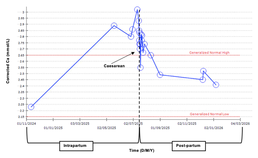
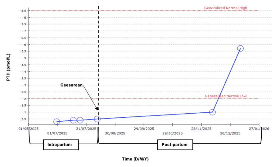
Figure 1: Peripartum trend in serum concentration of corrected calcium (mmol/L) and PTH (pmol/L)
- 1. Maekawa, A. S., Bennin, D., Hartery, S. A., Kirby, B. J., Poulton, I. J., St-Arnaud, R., Sims, N. A., & Kovacs, C. S. (2024). Maternal loss of 24-hydroxylase causes increased intestinal calcium absorption and hypercalcemia during pregnancy but reduced skeletal resorption during lactation in mice. Journal of Bone and Mineral Research, 39(12), 1793–1808. https://doi.org/10.1093/jbmr/zjae166
- 2. Sayers, J., Hynes, A. M., Srivastava, S., Dowen, F., Quinton, R., Datta, H. K., & Sayer, J. A. (2015). Successful treatment of hypercalcaemia associated with a CYP24A1 mutation with fluconazole. Clinical Kidney Journal, 8(4), 453–455. https://doi.org/10.1093/ckj/sfv028
- 3. CYP3A4 induction by rifampin: An alternative pathway for vitamin D inactivation in patients with CYP24A1 mutations. (2017). The Journal of Clinical Endocrinology & Metabolism, 102(?)*. https://pmc.ncbi.nlm.nih.gov/articles/PMC5443336/
- 4. Kovacs, C. S. (2016). Maternal mineral and bone metabolism during pregnancy, lactation, and post-weaning recovery. Physiological Reviews, 96(2), 449–547. https://doi.org/10.1152/physrev.00027.2015